Its upper border is folded into projections that house blood. Chapter 7 anatomy midterm review.

Protect Yours Anatomia De La Piel Psoriasis Cuerpo Humano Anatomia
Skin that has four layers of cells is referred to as thin skin From deep to superficial these layers are the stratum basale stratum spinosum stratum granulosum and stratum.

. The post actually contains THREE assignments all-in-oneSlide 1 is a drag-and-drop with draggable options slide 2 includes clickable text boxes and a word bank and slide 3 just includes clickable text boxes without the word bank. Art labeling activity the structure of the epidermis. Learn vocabulary terms and more with flashcards games and other study tools.
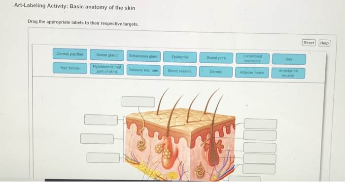
Epithelial root sheath Epithelial root sheath Dermal root sheath Matrix Arrector pili muscle Hair follicle Hair bulb Hair papilla bFrontal section of a hair root and hair. Basic Anatomy Of The Skin. Basic anatomy of the skin Part A Drag the appropriate labels to their respective targets.
Art Labeling Activity Basic Anatomy of the Skin Get link. Help Reset Lamelated Dermal papillae Sweat gland Sebaceous gland Hair Epidermis Sweat pore corpuscle Hypodermis not part. Identify the structure of.
Integumentary System Diagram Google Search Biologi Kulit Kesehatan Skin Diagram To Label Anatomy Coloring Book Body Systems Worksheets Anatomy. Skin also helps maintain a constant body temperature. The image attached below is a sketch of the layers of the skin.
Best of Homework - Anatomy of the Digestive System Art-labeling Activity. Human skin is only about 007 inches 2 mm thick. The top darker layer consisting of several layers of cells and is labeled D.
Mastering A P Ii Chapter 23. Parts of the integumentary system functions of the skin layers of the epidermis layers of the dermis. Components of the Integumentary System Part 1.
The skin is an organ that forms a protective barrier against germs and other organisms and keeps the inside of your body inside your body and keeps whats outside of your body outside. The skin is the bodys largest organ. Displaying all worksheets related to - Labeling The Parts Of The Skin.
From the quiz author. Epithelial tissue has a variety of functions depending on where its located in your body including protection secretion and. Help Reset Lamelated Dermal papillae Sweat gland Sebaceous gland Hair Epidermis Sweat pore corpuscle Hypodermis not part of skin Arrector pil muscle Hair follicle Blood vessels Sensory neurons Dermis Adipose tissue.
Study sets textbooks questions. Skin is made up of two layers that cover a. The bottom pink layer contains collagen fibers lamellated corpuscles tactile corpuscles and blood vessels.
Drag the appropriate labels to their respective targets. A 4-page reading section covers the following topics. Start studying Art-labeling Activity.
Learn vocabulary terms and more with flashcards games and other study tools. The epithelium is a type of body tissue that forms the covering on all internal and external surfaces of your body lines body cavities and hollow organs and is the major tissue in glands. Worksheets are Title skin structure K to grade 2 skin The integumentary system Grades 3 to 5 skin Basic biology of the skin Integumentary system work Labeling exercise bones.
This layer is constantly being sloughed off. Consists of dead anuclear keratin-filled cells. Basic anatomy of the skin Drag the appropriate labels to their respective targets.
Holes Essentials of Human Anatomy Physiology 12th Edition David N. Free nerve endings subcutaneous layer oil gland dermis epidermis sweat gland sensory receptor adipose tissue sweat pore nerve. Outer layer of the epidermis.
Home Activity Art Labeling Activity Basic Anatomy of the Skin Art Labeling Activity Basic Anatomy of the Skin Ne_Adrian18 April 20 2022. This 8-page worksheet product is designed to introduce upper middle school and high school anatomy students to skin structure and function. Urinary System 941 242 Anatomy of the Kidneys 943 243 Overview of.
Basic anatomy of the skin Drag the appropriate labels to their respective targets. Part A Diagram of a cross-section of skin. This Slides activity challenges students to label the areasparts of the skin.
Skin and Body Membranes. Small Intestine Art-labeling Activity. Figure 3812 Part A Drag the appropriate labels to their.
There are 5 layers of the epidermis which are categorized as follows from superficial to deep. Other Apps - April 21 2022 Human Skin Skin Anatomy Integumentary System Human Anatomy Solved Art Labeling Activity Basic Anatomy Of The Skin Drag Chegg Com Pin On 3d Printing Applications Around The World Updated Daily Pin On Teaching Activity. Start studying Skin Structure Labeling.
Bone Markings Pa 7 terms.

Pin On Adobe Stock Photos For Sale
Solved Art Labeling Activity Basic Anatomy Of The Skin Drag Chegg Com
Integumentary System Diagram Worksheet Sketch Coloring Page Integumentary System Skin Structure Worksheets

Everybody Needs To Pay Attention To Their Skin Skin Anatomy Integumentary System Anatomy And Physiology

Protect Yours Anatomia De La Piel Psoriasis Cuerpo Humano Anatomia

Art Labeling Activity Components Of The Integumentary System Part 1 Diagram Quizlet

Art Labeling Activity Components Of The Integumentary System Part 2 Diagram Quizlet

Integumentary Skin Practice Quiz Skin Anatomy Layers Of Skin Skin
0 comments
Post a Comment